38 bacterial cell picture with labels
Image Library | CDC Online Newsroom | CDC Under a high magnification of 21674X, this digitally-colorized, scanning electron microscopic (SEM) image depicts a view of a dividing, Escherichia coli bacterium, clearly displaying the point at which the bacteria's cell wall was splitting into two separate organisms. See PHIL 7137 for a black and white version of this image. Bacteria Labeled Diagram Stock Vector Image & Art - Alamy Download this stock vector: Bacteria Labeled Diagram - EG0XT7 from Alamy's library of millions of high resolution stock photos, illustrations and vectors.
50 Striking Microscopic Images of Viruses and Bacteria March 07, 2022. 1/50. Colorized transmission electron micrograph showing H1N1 influenza virus particles. Surface proteins on the virus particles are shown in black. (Credit: NIAID) Bacteria and ...

Bacterial cell picture with labels
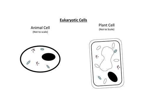
Animal Cell Labeled Pictures, Images and Stock Photos Browse 116 animal cell labeled stock photos and images available, or start a new search to explore more stock photos and images. Newest results Components of Eukaryotic cell, nucleus and organelles and plasma... Diagrams of animal and plant cells Labelled diagrams of typical animal and plant cells with editable layers. Golgi apparatus or Golgi body 600+ Free Bacteria & Virus Images - Pixabay Bacteria and virus high resolution images. Find your perfect picture for your project. 639 Free images of Bacteria / 7 ‹ › ... Structure of Bacterial Cell (With Diagram) - Biology Discussion It is a tough and rigid structure of peptidoglycan with accessory specific materials (e.g. LPS, teichoic acid etc.) surrounding the bacterium like a shell and lies external to the cytoplasmic membrane. It is 10-25 nm in thickness. It gives shape to the cell. Nucleus: The single circular double-stranded chromosome is the bacterial genome.
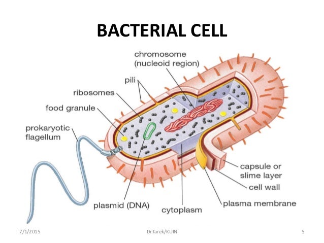
Bacterial cell picture with labels. Bacteria in Photos - photo gallery of bacteria facultatively anaerobic bacteria: Motility: nonmotile: Catalase test: catalase-positive: Oxidase test: negative* Spores: non-spore forming * Some species (non-human isolates) are positive: Streptococcus: ... (the cells stain a weak Gram-negative) Microscopic appearance: Spirochetes: Oxygen relationship: microaerobic: Motility: motile: Catalase ... Plant and Animal Cells - Labeled Graphics A compilation of plant and animal cell images with organelles and major structures labeled. Students can print images to help them learn the cell. ... if students missed the lab that day they can view a site with pictures to complete lab handout Plant Cell ... looks at cheek and onion cells. Prokaryote Coloring - color a typical bacteria cell ... 97,783 Bacteria Cell Stock Photos and Images - 123RF Bacteria Cell Stock Photos And Images 97,783 matches Page of 978 Structure of a bacterial cell. Anatomy of the prokaryote. unicellular organism. Vector diagram for your design, educational, medical, biological and science use Bacteria vector icon isolated on transparent background, Bacteria logo concept Bacterial Cell Structure Labeling Diagram | Quizlet Bacterial Cell Structure Labeling STUDY Learn Flashcards Write Spell Test PLAY Match Gravity Created by adkelly22 Terms in this set (16) Cytoplasm Water-based solution filling the entire cell Ribosomes Tiny particles composed of protein and RNA that are the sites of protein synthesis Nucleoid Composed of condensed DNA molecules. Cell Membrane
PHOTO GALLERY OF BACTERIA - Microbiology in pictures (the cells stain a weak Gram-negative) Microscopic appearance: Spirochetes: Oxygen relationship: microaerobic: Motility: motile: Catalase test:-Oxidase test:- ... Colonies of various bacteria. Bacteria photos. PICTURE OF THE MONTH. BACTERIA 2013 DECEMBER. DISK DIFFUSION METHOD FOR TESTING OF ANTIBIOTIC SUSCEPTIBILITY OF BACTERIA: Bacteria Cell Structures with Labels Stock Vector - Dreamstime Get 15 images free trial Bacteria Cell Structures with labels Royalty-Free Vector Bacterial cell structures labeled on a bacillus cell with nucleoid DNA and ribosomes. External structures include the capsule, pili, and flagellum. Morphology of internal structures of bacteria. cell anatomy bacteria, prokaryotic cell, cell, internal structures, Label the Bacterium Cell - EnchantedLearning.com flagellum - A long whip-like structure used for locomotion (movement). Some bacteria have more than one flagellum. pili - (singular is pilus) Hair-like projections that allow bacterial cells to stick to surfaces and transfer DNA to one another. plasma membrane - A permeable membrane located within the cell wall. Bacteria in Microbiology - shapes, structure and diagram Bacterial endospores layers Bacteria cells are the smallest living cells that are known; even though viruses are smaller than bacteria, viruses are not living cells. There are different types of bacteria with various sizes, shapes, and structures. The bacteria shapes, structure, and labeled diagrams are discussed below. Sizes
3 Common Bacteria Shapes - ThoughtCo Bacteria Shapes The three basic shapes of bacteria include cocci (blue), bacilli (green), and spirochetes (red). PASIEKA/Science Photo Library/Getty Images By Regina Bailey Updated on August 20, 2019 Bacteria are single-celled, prokaryotic organisms that come in different shapes. Bacteria (Prokaryote) Cell Coloring - The Biology Corner Color the cell wall purple. 2. On the inside of the cell wall is the cell membrane . Its job is to regulate what comes in and out of the cell. Color the cell membrane pink. 3. The surface of some bacteria cells is covered in pilus, which help the cell stick to surfaces. Color the pilus light green. 4. Bacterial Cell Structure Diagram - Quizlet PLAY. Bacteria. Domain of unicellular prokaryotes that have cell walls containing peptidoglycan. Flagella. a slender whip-like structure that enables the bacteria to propel itself; an organ of locomotion. Ribosome. a cytoplasmic nuclear protein that is required in mRNA translation and protein synthesis. Nucleoid. Bacteria Labeled Stock Illustrations - 225 Bacteria Labeled Stock ... Bacteria Labeled Stock Illustrations - 225 Bacteria Labeled Stock Illustrations, Vectors & Clipart - Dreamstime Bacteria Labeled Illustrations & Vectors Most relevant Best selling Latest uploads Within Results People Pricing License Media Properties More Safe Search 225 bacteria labeled illustrations & vectors are available royalty-free. Next page
Plant Cell Labeled Pictures, Images and Stock Photos Labeled bacteria internal structure scheme Cyanobacteria vector illustration. Labeled educational bacteria internal structure scheme. Biological blue green algae diagram with carboxysome, thylakoid and phycobilisome parts location inside cell. plant cell labeled stock illustrations ... Data collection during medical research plant cell labeled ...
Structure of Bacteria (With Diagram) | Microbiology Bacterial cell wall is extremely thin (10-25 nm thick) and provides rigidity and a definite shape to the cell. 7. Chemically, the cell wall is composed of mucopeptide (murein) scaffolding or platform formed by N- acetyl glucosamine and N-acetyl muramic acid molecules arranged in alternate chains. 8.
Structure of a bacterial cell, labeled. Stock Illustration Download Structure of a bacterial cell, labeled. Stock Illustration and explore similar illustrations at Adobe Stock. Adobe Stock. Photos Illustrations Vectors Videos Audio Templates Free Premium Editorial Fonts. Plugins. 3D. Photos Illustrations Vectors Videos Audio Templates Free Premium Editorial Fonts.
Bacterial Cell Structures Labeled On Bacillus Stock ... - Shutterstock Stock Vector ID: 1522904069 Bacterial cell structures labeled on a bacillus cell with nucleoid DNA and ribosomes. External structures include the capsule, pili, and flagellum. Vector Formats EPS 2789 × 2200 pixels • 9.3 × 7.3 in • DPI 300 • JPG Show more Vector Contributor O OSweetNature Similar images See all Assets from the same collection
how to draw & label bacteria - YouTube | Science diagrams, Teaching, Labels The cytoplasm of prokaryotic cells lacks in well defined cell organelles such as endoplasmic reticulum, Golgi apparatus, Mitochondria,centrioles, nucleoli, cytoskeleton. The size of these cells range between 1 micrometre to 3 micrometres, so they are barely visible under the light microscope. Now let's moveon to diagram. 1.Draw a capsule shape… S
Different Size, Shape and Arrangement of Bacterial Cells When viewed under light microscope, most bacteria appear in variations of three major shapes: the rod (bacillus), the sphere (coccus) and the spiral type (vibrio). In fact, structure of bacteria has two aspects, arrangement and shape. So far as the arrangement is concerned, it may Paired (diplo), Grape-like clusters (staphylo) or Chains (strepto).
Bacterial Staining Microbiology Images ... - Science Prof Online 1. Endospore stain of Bacillus subtilis showing both endospores (green) & vegetative cells (pink) @1000xTM; 2. Negative endospore stain showing only vegetative cells @1000xTM; 3. Malachite green primary staining step of endopore stain with slide being heated over water bath; 4. Applying counterstain (safrinin) to bacterial smear as last step of endospore stain; Endospore stained slide, with ...
Interactive Bacteria Cell Model - CELLS alive The three primary shapes in bacteria are coccus (spherical), bacillus (rod-shaped) and spirillum (spiral). Mycoplasma are bacteria that have no cell wall and therefore have no definite shape. Outer Membrane: This lipid bilayer is found in Gram negative bacteria and is the location of lipopolysaccharide (LPS) in these bacteria.
Bacterial cells - Cell structure - Edexcel - GCSE Combined Science ... Feature Eukaryotic cell (plant and animal cell) Prokaryotic cell (bacterial cell) Size: Most are 5 μm - 100 μm: Most are 0.2 μm - 2.0 μm: Outer layers of cell

166 best images about Microbiology and Microbiology Jokes to share on Pinterest | Biology, Blood ...
2,284 Animal cell labeled Images, Stock Photos & Vectors - Shutterstock 2,277 animal cell labeled stock photos, vectors, and illustrations are available royalty-free. See animal cell labeled stock video clips Image type Orientation Color People Artists More Sort by Biology Animals and Wildlife Healthcare and Medical Cooking Software cell eukaryote in vitro experiment cell culture Turn on AI Powered Search
Bacteria - Definition, Structure, Diagram, Classification The bacteria diagram given below represents the structure of bacteria with its different parts. The cell wall, plasmid, cytoplasm and flagella are clearly marked in the diagram. Bacteria Diagram representing the Structure of Bacteria Structure of Bacteria The structure of bacteria is known for its simple body design.
Structure of Bacterial Cell (With Diagram) - Biology Discussion It is a tough and rigid structure of peptidoglycan with accessory specific materials (e.g. LPS, teichoic acid etc.) surrounding the bacterium like a shell and lies external to the cytoplasmic membrane. It is 10-25 nm in thickness. It gives shape to the cell. Nucleus: The single circular double-stranded chromosome is the bacterial genome.
600+ Free Bacteria & Virus Images - Pixabay Bacteria and virus high resolution images. Find your perfect picture for your project. 639 Free images of Bacteria / 7 ‹ › ...
Animal Cell Labeled Pictures, Images and Stock Photos Browse 116 animal cell labeled stock photos and images available, or start a new search to explore more stock photos and images. Newest results Components of Eukaryotic cell, nucleus and organelles and plasma... Diagrams of animal and plant cells Labelled diagrams of typical animal and plant cells with editable layers. Golgi apparatus or Golgi body


Post a Comment for "38 bacterial cell picture with labels"